Mri Of Normal Brain Photograph by Living Art Enterprises Fine Art America
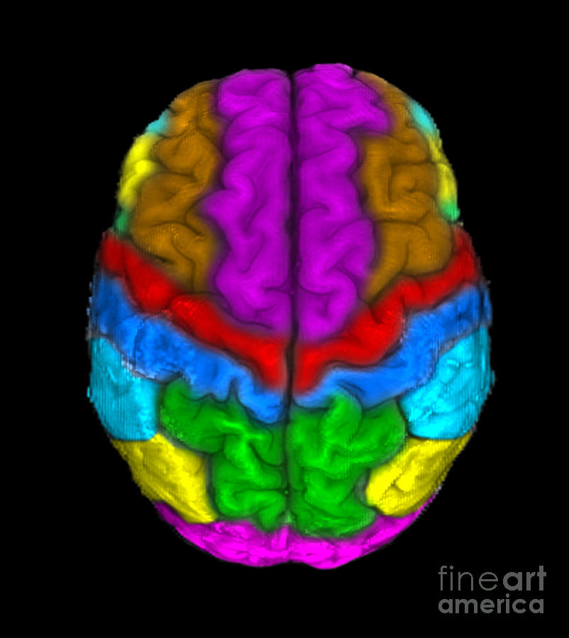
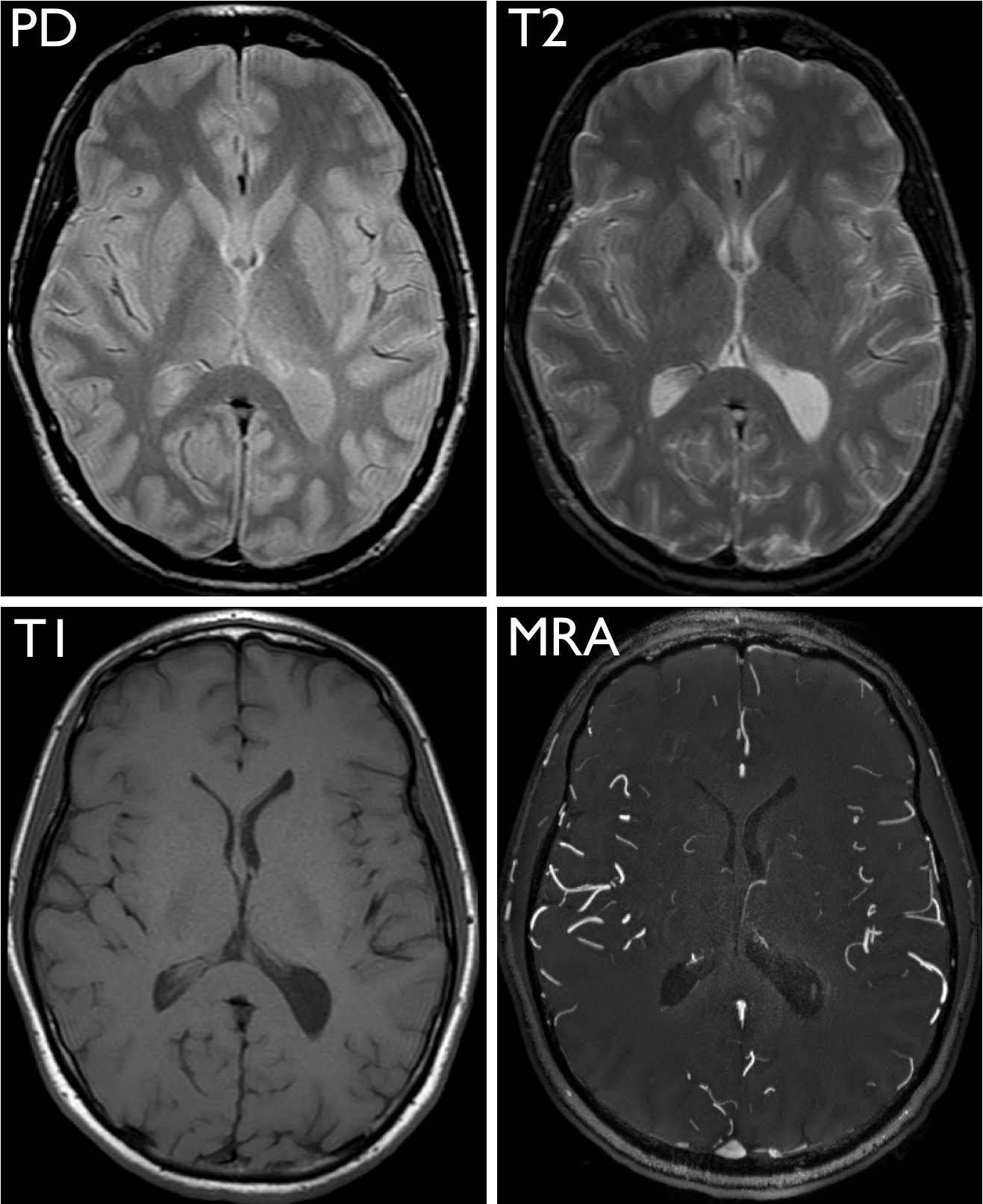
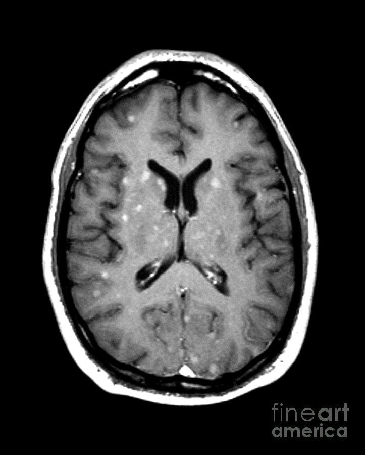
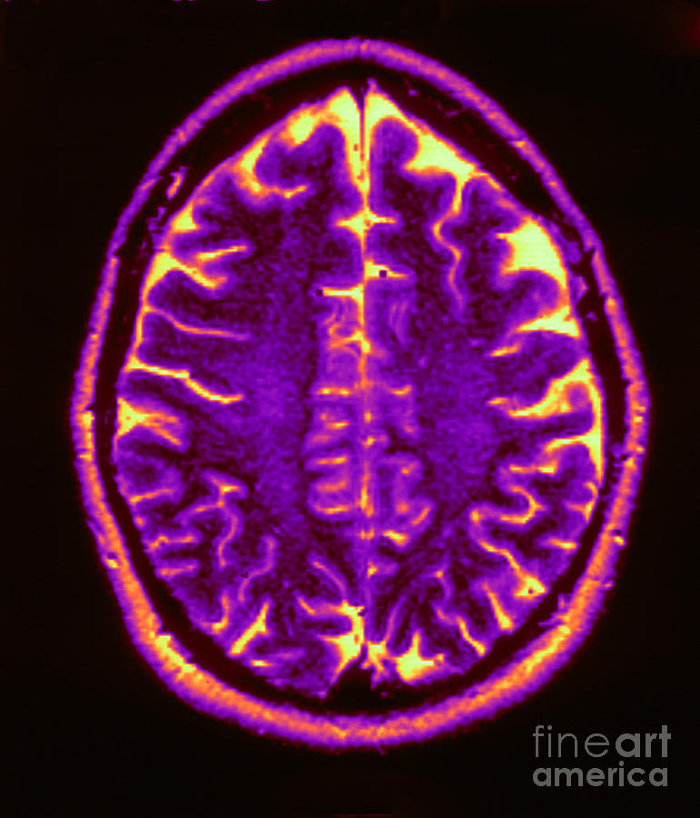
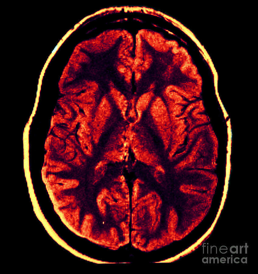
Normal brain MRI A brain MRI is one of the most commonly performed techniques of medical imaging. It enables clinicians to focus on various parts of the brain and examine their anatomy and pathology, using different MRI sequences, such as T1w, T2w, or FLAIR.
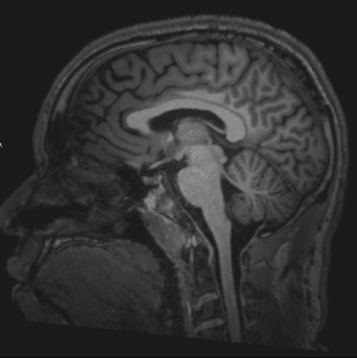
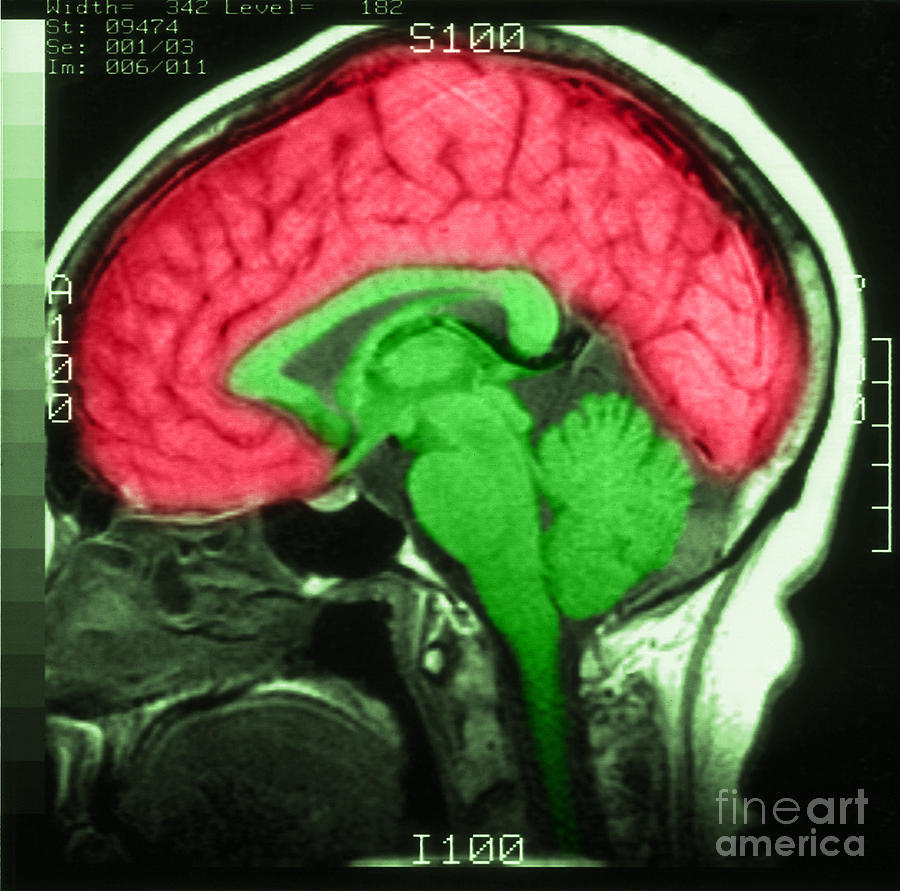
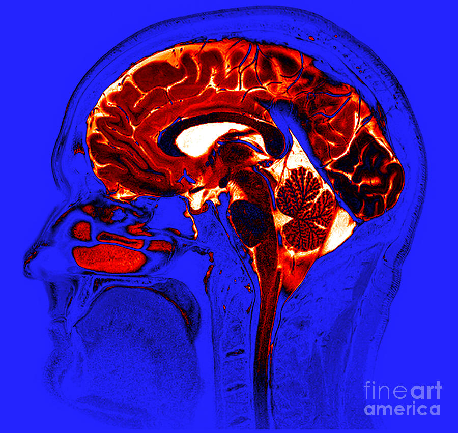
Image Normal Brain MRI (Sagittal) Slide 3 MSD诊疗手册专业版
EEG (electroencephalogram): An electroencephalogram (EEG) is a test that detects electrical activity in your brain using small, flat metal discs (electrodes) attached to your scalp. Your brain cells communicate via electrical impulses and are active all the time, even when you're asleep. This activity shows up as wavy lines on an EEG recording.
Exploring the Brain How Are Brain Images Made with MRI? UCSF Radiology
Trauma can damage your brain tissue, neurons, and nerves. This damage affects your brain's ability to communicate with the rest of your body. Examples of brain injuries include: hematomas. blood.
Mri Of Normal Brain Photograph by Science Source Fine Art America
Young Frankenstein Abby Normal Aesir Books 664 subscribers Subscribe Subscribed 725K views 7 years ago More clips at our blog http://www.aesirbooks-eu.s3dsdesign.com This trailer is used under.
Abnormal Mri Of Brain Photograph by Medical Body Scans
Brain abnormalities are wide ranging and can be both organic, developmental or a combination of both in origin. Research has linked the presence of brain abnormalities to a variety of conditions including developmental disorders such as Autism [], Schizophrenia [], Alcoholism [], various types of brain tumors, and dementias.With the remarkably complex nature of early brain development, human.
Mri Of Normal Brain Photograph by Science Source Fine Art America
These antibodies disrupt normal brain signaling and cause brain swelling, or encephalitis. It can affect both men and women, however is more common among women. It primarily affects the young, including children and young adults. Some patients also have a tumor associated with this disease; the most common type is an ovarian teratoma in women.
Mri Of Normal Brain Photograph by Science Source Fine Art America
Young Frankenstein 1974-The brain came from Abbey Normal - YouTube 0:00 / 1:38 Igor is being questioned about where he got the brain for the monster. He has a great answer for a very funny.
Brain MRI How to read MRI brain scan Kenhub
The Aβ is a 4 kDa fragment of the amyloid precursor protein (APP), a larger precursor molecule widely produced by brain neurons, vascular and blood cells (including platelets), and, to a lesser.
Normal Brain, Mri Photograph by Living Art Enterprises Fine Art America
Promising to not get angry if Igor confesses to his mistake, he eventually coaxes out the truth: that the brain came from someone named "Abby Normal." The doctor quickly realizes he's placed an.
Abnormal brain interactions harm consciousness
Lesson Transcript. Ashli has a Master's Degree in Biology and has taught biology at different grade levels including college, elementary, and middle school. Brain abnormalities occur when vital.
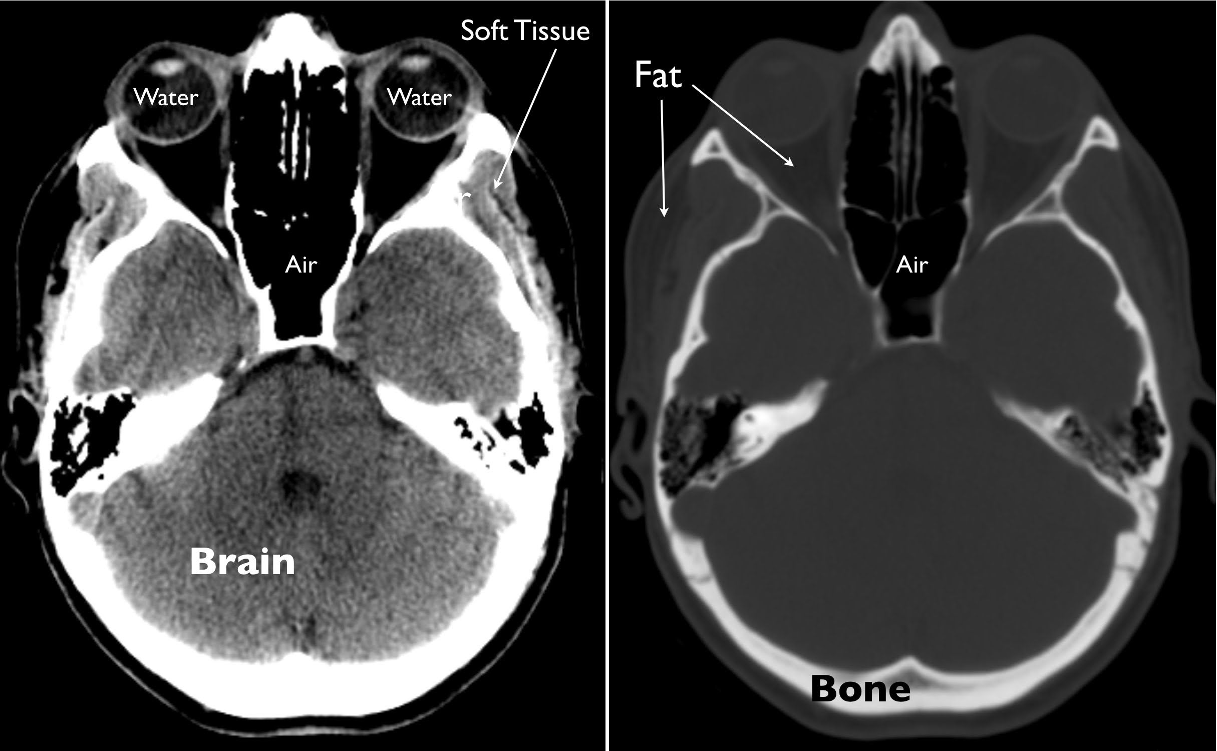
Exploring the Brain How Are Brain Images Made with CT? UCSF Radiology
Igor: "Abby Normal. Yes, I am almost sure it was Abby Normal." Frankenstein: "Are you telling me that I put an abnormal brain into a 7-foot-long, 54-inch-wide gorilla!!!"
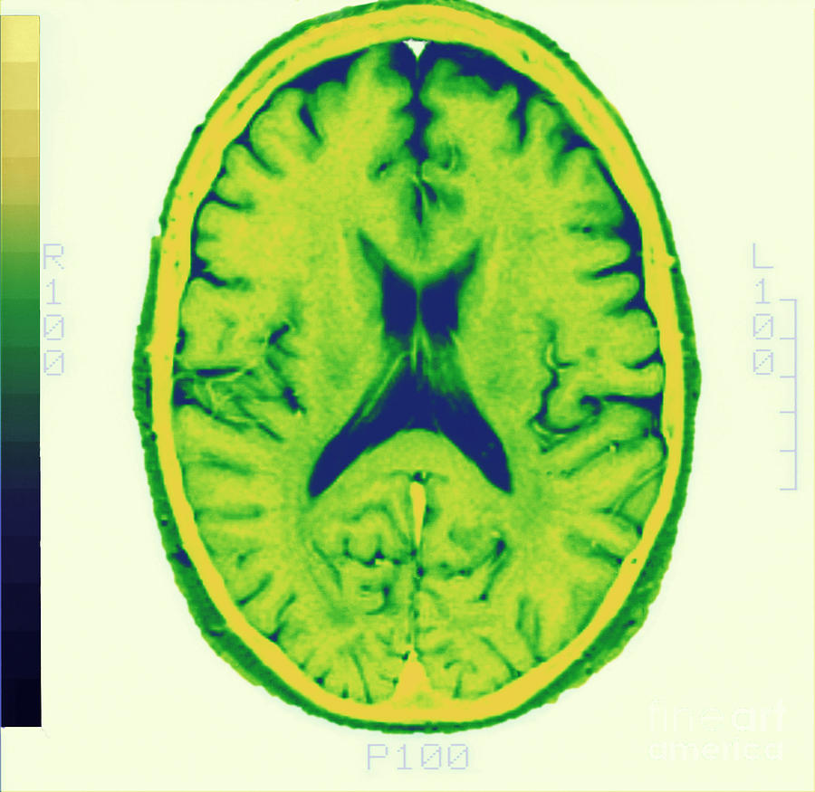
Normal Brain Photograph by Zephyr/science Photo Library Fine Art America
The cerebellum makes up approximately 10% of the brain's total size, but it accounts for more than 50% of the total number of neurons located in the entire brain. The cerebellum is comprised of small lobes and serves several functions. It receives information from the inner ear's balance system, sensory nerves, and auditory and visual systems.
Frontiers Abnormal Brain Structure Morphology in EarlyOnset
Frontal lobe. Your frontal lobe is at the front of your head. Lesions in your frontal lobe can lead to certain symptoms or conditions, including: Trouble with learning. Visual-motor function. Executive dysfunction and problems with attention (planning, focusing and inhibition). Agitation and mood swings.
Mri Of Normal Brain Photograph by Science Source Fine Art America
In an MRI report, the white spots might be described as: "High signal intensity areas". "White matter hyperintensities" (lesions that appear bright white on certain sequences of MRI scans) " Leukoaraiosis " (a term that is used if the spots are thought to be caused by decreased blood flow. "Nonspecific white matter changes".
Mri Of Normal Brain Photograph by Science Source Fine Art America
(I apologize for the display and the sepia-like tone of the video. I have no clue why it looks this way and I can't get it to look normal no matter what I t.

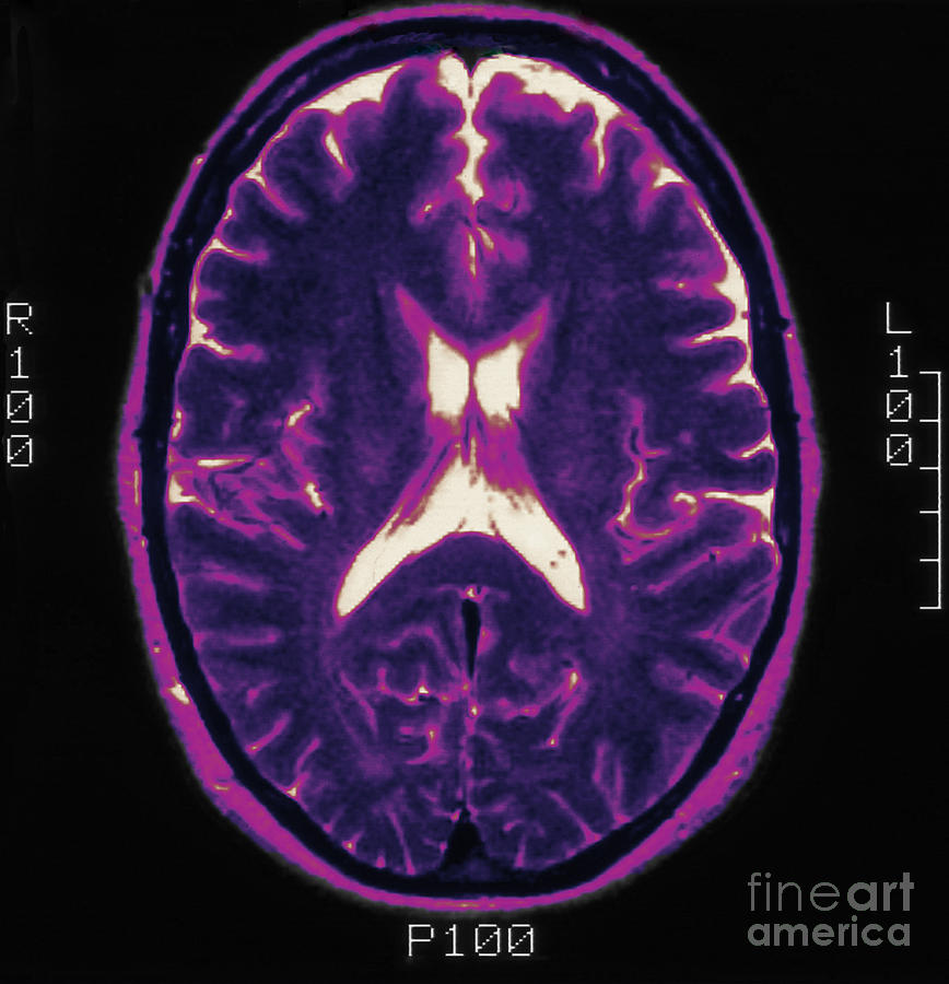
MRI image of the brain in an axial view showing the “precontrast FLAIR
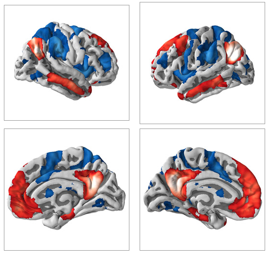
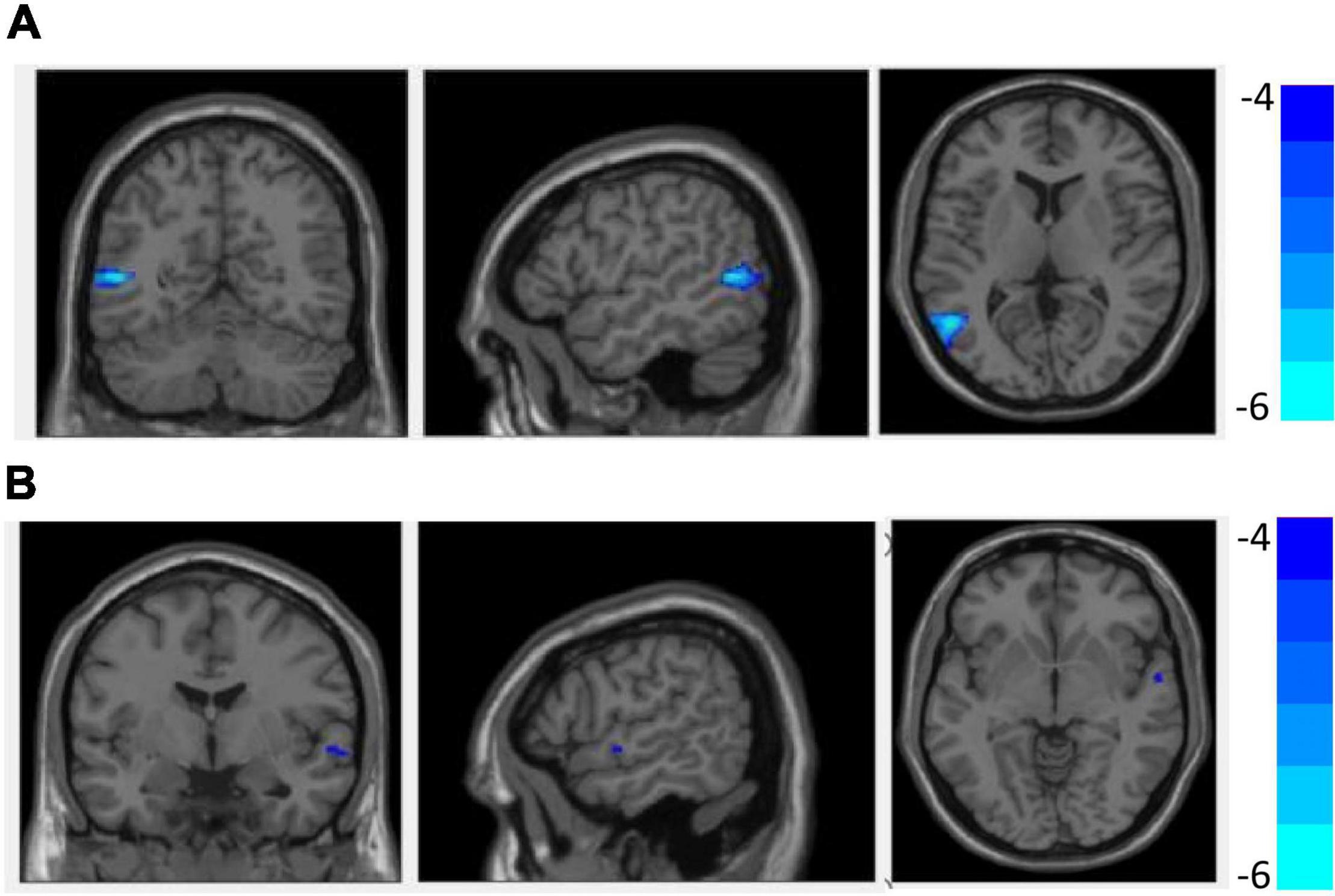
Our results suggested that abnormal neural activity in these brain regions may represent a potential neurobiological diathesis or predisposition to suicidal behavior in youth depression. Keywords: Amplitude of low frequency fluctuation; Functional magnetic resonance imaging (fMRI); Impulsivity; Resting state; Suicide attempt; Youth depression.
